In the last several decades, medical technology has advanced in leaps and bounds. One of the most advanced technological instruments used in medicine today is the nuclear bone scan. A nuclear bone scan uses a radioactive tracer and high-resolution camera to identify areas of new bone growth or areas in which bone has broken down.
For What Purpose is a Nuclear Bone Scan Used?
There are several ways that a nuclear bone scan can be used. In general, bone scans are an excellent tool for identifying and evaluating damage to bones, assessing infections and trauma, and detecting cancer that has metastasized to the bones. Metastasize is a term used to define the spread of cancer. It should be noted that other tools such as X-rays can be used in the above situations, but nuclear bone scans are usually more efficient at detecting issues in advance. In some cases, a nuclear bone scan can detect problems months in advance of an X-ray, while other issues are usually limited to only a few days.
The Nuclear Bone Scan Process
The nuclear bone scan procedure is not invasive and is, for the most part, painless. The nuclear bone scan process employs a radioactive tracer substance that is normally injected into a vein in one of the patient’s arms. A high-resolution camera scans the body and picks up specific radioactive substances.
Preparing for a Nuclear Bone Scan
A nuclear bone scan is usually done at a hospital or at a special center. Preparing for a nuclear bone scan is quite easy. Before arriving for the scan, do not drink any liquids for about 4 hours. In addition, right before the tracer is inserted into the patient’s arm, the doctor will asked the patient to go to the bathroom.
During the Process
A nuclear medicine technician usually administers a nuclear bone scan with the help of the nursing staff. When they are ready to insert the tracer, they will check that the patient has not drunk any fluids and has recently gone to the bathroom. To insert the tracer, they will simply inject the tracer substance into a vein in the arm. It is a simple procedure, and the patient should only feel a slight pinch.
The tracer substance is slightly radioactive, which will allow it to show up on the scanning device (camera) and help the radiologist or doctor to evaluate the patient’s condition. As the tracer substance is inserted into the body, it travels throughout the bloodstream, coming into contact with all the patient’s bones.
Once the tracer substance is inserted, it usually takes up to 3 hours to fully circulate throughout the body. During this time, the patient can relax, read a book, etc. In addition, the patient is usually asked to drink about 4 to 6 glasses of water, which helps remove excess radioactive waste from his/her body.
Immediately before the scanning process begins, the patient will once again be asked to go to the bathroom to eliminate any waste. Urine in the bladder may obstruct the scanning of specific bones in the pelvic area.
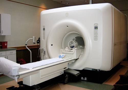
The scanning device consists of a large scanning camera that is placed very close to the body. The patient will be asked to undress fully or partially and to remove any jewelry he/she may be wearing. Once undressed, the patient is usually given a blanket or simple paper robe.
Before the scan is done, the patient has to lie down on his/her back on a table. After lying down, he/she will be asked to be very still. Any movement may blur the images taken. Besides lying on his/her back, the technician might ask the patient to change position to get more detailed images of specific areas of his/her body. It should be noted that the scanner does not emit any radiation. The actual scanning process is usually not time consuming or painful and most scans are completed within an hour’s time. It should be noted that a bone scan may be for the entire body or for just a specific body part.
Radiation is eliminated from the body in just a day or two via the natural processes of waste elimination. All the patient has to do is go to the bathroom as usual. The amount of radiation in the tracer is very small, so no risk is posed to others.
Results from a Nuclear Bone Scan
The bones absorb the tracer element that is inserted into the arm. The amount of tracer the bones absorbed could indicate abnormalities. For instance, uniform absorption is usually a sign that there are no abnormalities.
If only a little or no tracer was absorbed, it could be due to lack of blood flow. The term used for low blood flow to the bone is bone infarction. A bone infarction may mean that cancer is present.
Areas with a bright spot, indicating a lot of absorption, may indicate that this area of the bone has an infection, a fracture, tumor, or a certain type of cancer. Whatever the case may be, the doctor and radiologist will go over the results and discuss them with the patient.


bridge
how do they do a bone scan on my wrist. do i have to go in all the way in because when i had the mri i went in half way.
Leslie witten
I have recently had a MRI done on my lower back area as well as a number of blood tests. The blood tests were a success with no problems. The MRI scan indicated that there was nothing conclusive to the doctors findings. I was then told that I must undergo a bone scan to ascertain whether there is arthritis present. Why is it now necessary to have this scan done when surely a MRI would be sufficient. At present I am experiencing a lot of pain in the lumber area with a certain amount of muscle spasms in that area. Also have not had any proper sleep recently.
Mike
MRI scans measure water content and therefore detect tissue best. Bone scans and x-rays detect bones best.
Shirley
I just had a routine blood lab test done. The results showed I had an elevated emzyme 170 Alkaline Phosphate. Is this a real alarming. I was told that I needed a bone scan. It was also stated that the emzyme elevation causes bones to emit. I’m not sure what this means.